GV

GeezerVison

The World Wide Web, restored to 800 pixels of glory.

Home|About|Gaming cabinet|Archives|How it works|Starter sites

On this site

  • Search
  • Advanced Search
  • Sequence Similarity Search
  • 3D Similarity Search
  • Chemical Similarity
  • Browse Annotations
  • New Entries
  • Unreleased Entries
  • PDB Statistics
  • Tools at-a-glance
  • Mol* (MolStar)
  • Sequence Annotations Viewer
  • Sequence Annotations in 3D
  • Genome View

This view

  • Original page
  • New address

GeezerVison is showing www.rcsb.org — RCSB PDB - 1IIL: CRYSTAL STRUCTURE OF PRO253ARG APERT MUTANT FGF RECEPTOR 2 (FGFR2) IN COMPLEX WITH FGF2

  • Structure Summary
  • Structure
  • Annotations
  • Experiment
  • Sequence
  • Genome
  • Versions

  • FASTA Sequence
  • mmCIF Format
  • mmCIF Format (Header)
  • Legacy PDB Format
  • Legacy PDB Format (Header)
  • FASTA Sequence
  • PDBx/mmCIF Format
  • PDBx/mmCIF Format (gz)
  • BinaryCIF Format (gz)
  • Legacy PDB Format
  • Legacy PDB Format (gz)
  • PDBML/XML Format (gz)
  • Validation Full (PDF - gz)
  • Validation (XML - gz)
  • Validation (CIF - gz)
  • Biological Assembly 1 (CIF - gz) 
  • Biological Assembly 2 (CIF - gz) 
  • Biological Assembly 3 (CIF - gz) 
  • Biological Assembly 4 (CIF - gz) 
  • Biological Assembly 1 (PDB - gz)
  • Biological Assembly 2 (PDB - gz)
  • Biological Assembly 3 (PDB - gz)
  • Biological Assembly 4 (PDB - gz)
 Data API 

 1IIL | pdb_00001iil

CRYSTAL STRUCTURE OF PRO253ARG APERT MUTANT FGF RECEPTOR 2 (FGFR2) IN COMPLEX WITH FGF2

  • PDB DOI: https://doi.org/10.2210/pdb1IIL/pdb
  • Classification: GROWTH FACTOR/GROWTH FACTOR RECEPTOR
  • Organism(s): Homo sapiens
  • Expression System: Escherichia coli BL21(DE3)
  • Mutation(s): Yes 

  • Deposited: 2001-04-23 Released: 2001-05-09 
  • Deposition Author(s): Ibrahimi, O.A., Eliseenkova, A.V., Plotnikov, A.N., Ornitz, D.M., Mohammadi, M.

Experimental Data Snapshot

  • Method: X-RAY DIFFRACTION
  • Resolution: 2.30 Å
  • R-Value Free: 
    0.259 (Depositor) 
  • R-Value Work: 
    0.237 (Depositor) 
  • R-Value Observed: 
    0.237 (Depositor) 

Starting Model: experimental
View more details

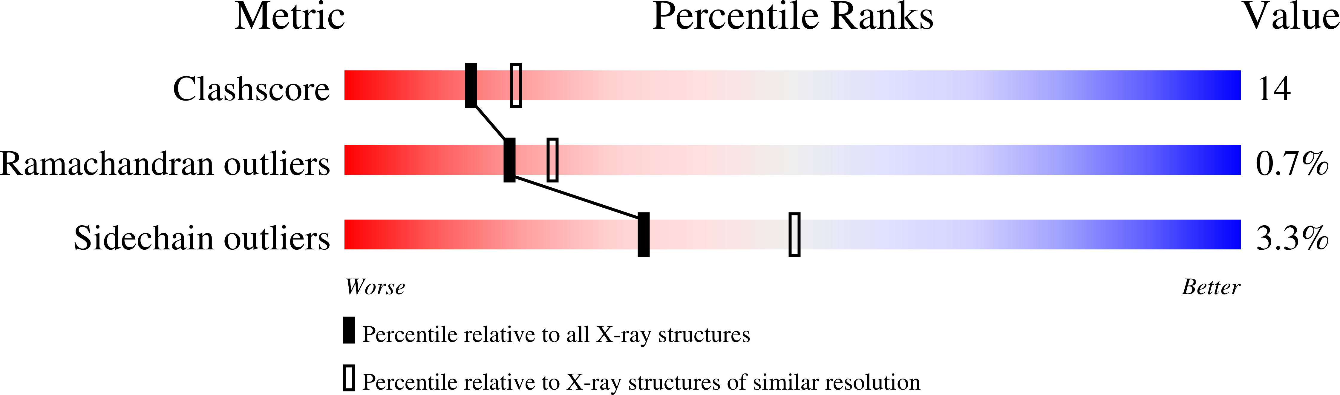
wwPDB Validation 3D Report Full Report

Validation slider image for 1IIL

This is version 1.5 of the entry. See complete history. 

Literature

  • Download Mendeley

Structural basis for fibroblast growth factor receptor 2 activation in Apert syndrome.

Ibrahimi, O.A., Eliseenkova, A.V., Plotnikov, A.N., Yu, K., Ornitz, D.M., Mohammadi, M.

(2001) Proc Natl Acad Sci U S A 98: 7182-7187

  • PubMed: 11390973 Search on PubMedSearch on PubMed Central
  • DOI: https://doi.org/10.1073/pnas.121183798
  • Primary Citation Related Structures: 
    1II4, 1IIL

  • PubMed Abstract: 

    Apert syndrome (AS) is characterized by craniosynostosis (premature fusion of cranial sutures) and severe syndactyly of the hands and feet. Two activating mutations, Ser-252 --> Trp and Pro-253 --> Arg, in fibroblast growth factor receptor 2 (FGFR2) account for nearly all known cases of AS. To elucidate the mechanism by which these substitutions cause AS, we determined the crystal structures of these two FGFR2 mutants in complex with fibroblast growth factor 2 (FGF2). These structures demonstrate that both mutations introduce additional interactions between FGFR2 and FGF2, thereby augmenting FGFR2-FGF2 affinity. Moreover, based on these structures and sequence alignment of the FGF family, we propose that the Pro-253 --> Arg mutation will indiscriminately increase the affinity of FGFR2 toward any FGF. In contrast, the Ser-252 --> Trp mutation will selectively enhance the affinity of FGFR2 toward a limited subset of FGFs. These predictions are consistent with previous biochemical data describing the effects of AS mutations on FGF binding. Alterations in FGFR2 ligand affinity and specificity may allow inappropriate autocrine or paracrine activation of FGFR2. Furthermore, the distinct gain-of-function interactions observed in each crystal structure provide a model to explain the phenotypic variability among AS patients.


  • Organizational Affiliation: 
    • Department of Pharmacology, New York University School of Medicine, New York, NY 10016, USA.
Asymmetric Unit

 Explore in 3D: Structure | Sequence Annotations | Validation Report

Biological Assembly 1  

 Explore in 3D: Structure | Sequence Annotations | Validation Report


Global Symmetry: Asymmetric - C1 
Global Stoichiometry: Hetero 2-mer - A1B1 


Find Similar Assemblies

Biological assembly 1 assigned by authors and generated by PISA (software)

Biological Assembly 2  

 Explore in 3D: Structure | Sequence Annotations | Validation Report


Global Symmetry: Asymmetric - C1 
Global Stoichiometry: Hetero 2-mer - A1B1 


Find Similar Assemblies

Biological assembly 2 assigned by authors and generated by PISA (software)

Biological Assembly 3  

 Explore in 3D: Structure | Sequence Annotations | Validation Report


Global Symmetry: Asymmetric - C1 
Global Stoichiometry: Hetero 2-mer - A1B1 


Find Similar Assemblies

Biological assembly 3 assigned by authors and generated by PISA (software)

Biological Assembly 4  

 Explore in 3D: Structure | Sequence Annotations | Validation Report


Global Symmetry: Asymmetric - C1 
Global Stoichiometry: Hetero 2-mer - A1B1 


Find Similar Assemblies

Biological assembly 4 assigned by authors and generated by PISA (software)

Macromolecule Content 

  • Total Structure Weight: 167.8 kDa 
  • Atom Count: 10,677 
  • Modeled Residue Count: 1,361 
  • Deposited Residue Count: 1,500 
  • Unique protein chains: 2

Macromolecules

Find similar proteins by:
  • 100%
  • 95%
  • 90%
  • 80%
  • 70%
  • 60%
  • 50%
  • 40%
  • 30%
|  3D Structure
Entity ID: 1
MoleculeChains  Sequence LengthOrganismDetailsImage
HEPARIN-BINDING GROWTH FACTOR 2
A, B, C, D
155Homo sapiensMutation(s): 2 
UniProt & NIH Common Fund Data Resources
Find proteins for P09038 (Homo sapiens)
Explore P09038 
Go to UniProtKB:  P09038
GTEx:  ENSG00000138685 
Entity Groups
Sequence Clusters30% Identity50% Identity70% Identity90% Identity95% Identity100% Identity
UniProt GroupP09038
Sequence Annotations
Expand
Reference Sequence
Find similar proteins by:
  • 100%
  • 95%
  • 90%
  • 80%
  • 70%
  • 60%
  • 50%
  • 40%
  • 30%
|  3D Structure
Entity ID: 2
MoleculeChains  Sequence LengthOrganismDetailsImage
FIBROBLAST GROWTH FACTOR RECEPTOR 2
E, F, G, H
220Homo sapiensMutation(s): 1 
EC: 2.7.1.112 (PDB Primary Data), 2.7.10.1 (UniProt)
UniProt & NIH Common Fund Data Resources
Find proteins for P21802 (Homo sapiens)
Explore P21802 
Go to UniProtKB:  P21802
GTEx:  ENSG00000066468 
Entity Groups
Sequence Clusters30% Identity50% Identity70% Identity90% Identity95% Identity100% Identity
UniProt GroupP21802
Sequence Annotations
Expand
Reference Sequence

Experimental Data & Validation

Experimental Data

  • Method: X-RAY DIFFRACTION
  • Resolution: 2.30 Å
  • R-Value Free:  0.259 (Depositor) 
  • R-Value Work:  0.237 (Depositor) 
  • R-Value Observed: 0.237 (Depositor) 
Space Group: P 1
Unit Cell:
Length ( Å )Angle ( ˚ )
a = 70.796α = 90.01
b = 72.49β = 89.82
c = 89.919γ = 90
Software Package:
Software NamePurpose
DENZOdata reduction
SCALEPACKdata scaling
AMoREphasing
CNSrefinement

Structure Validation

View Full Validation Report


View more in-depth experimental data

Entry History 

Deposition Data

  • Released Date: 2001-05-09 
  • Deposition Author(s): Ibrahimi, O.A., Eliseenkova, A.V., Plotnikov, A.N., Ornitz, D.M., Mohammadi, M.

Revision History  (Full details and data files)

  • Version 1.0: 2001-05-09
    Type: Initial release
  • Version 1.1: 2008-04-27
    Changes: Version format compliance
  • Version 1.2: 2011-07-13
    Changes: Version format compliance
  • Version 1.3: 2021-10-27
    Changes: Database references
  • Version 1.4: 2023-08-16
    Changes: Data collection, Refinement description
  • Version 1.5: 2024-11-13
    Changes: Structure summary
You are visitor 01402503 — GeezerVison does not store the pages you look up.
Best viewed with Netscape Navigator 4.7 or Internet Explorer 6 at 800×600. No cookies required.