GeezerVisonThe World Wide Web, restored to 800 pixels of glory. | |||||||||||||||||||||||||||||||||||||||||||||||||||||||||||||||||||||||||||||||||||||||||||||||||||||||||
| Home|About|Gaming cabinet|Archives|How it works|Starter sites | |||||||||||||||||||||||||||||||||||||||||||||||||||||||||||||||||||||||||||||||||||||||||||||||||||||||||
GeezerVison is showing www.rcsb.org — RCSB PDB - 2XSI: Human serum albumin complexed with dansyl-L-glutamate and myristic acid
| |||||||||||||||||||||||||||||||||||||||||||||||||||||||||||||||||||||||||||||||||||||||||||||||||||||||||
Entity ID: 1 | |||||
|---|---|---|---|---|---|
| Molecule | Chains | Sequence Length | Organism | Details | Image |
| SERUM ALBUMIN | 585 | Homo sapiens | Mutation(s): 0 |  | |
UniProt & NIH Common Fund Data Resources | |||||
GTEx: ENSG00000163631 | |||||
Entity Groups | |||||
| Sequence Clusters | 30% Identity50% Identity70% Identity90% Identity95% Identity100% Identity | ||||
| UniProt Group | P02768 | ||||
Sequence AnnotationsExpand | |||||
Reference Sequence | |||||
Small Molecules
| Ligands 2 Unique | |||||
|---|---|---|---|---|---|
| ID | Chains | Name / Formula / InChI Key | 2D Diagram | 3D Interactions | |
| 9NE Download:Ideal Coordinates CCD File | H [auth A], I [auth A] | DANSYL-L-GLUTAMATE C17 H20 N2 O6 S OXWKCHDQOLCMPE-ZDUSSCGKSA-N |  | Interactions Interactions & Density | |
| MYR Download:Ideal Coordinates CCD File
| B [auth A] C [auth A] D [auth A] E [auth A] F [auth A] B [auth A], C [auth A], D [auth A], E [auth A], F [auth A], G [auth A] | MYRISTIC ACID C14 H28 O2 TUNFSRHWOTWDNC-UHFFFAOYSA-N |  | Interactions | |
Experimental Data & Validation
Experimental Data
- Method: X-RAY DIFFRACTION
- Resolution: 2.70 Å
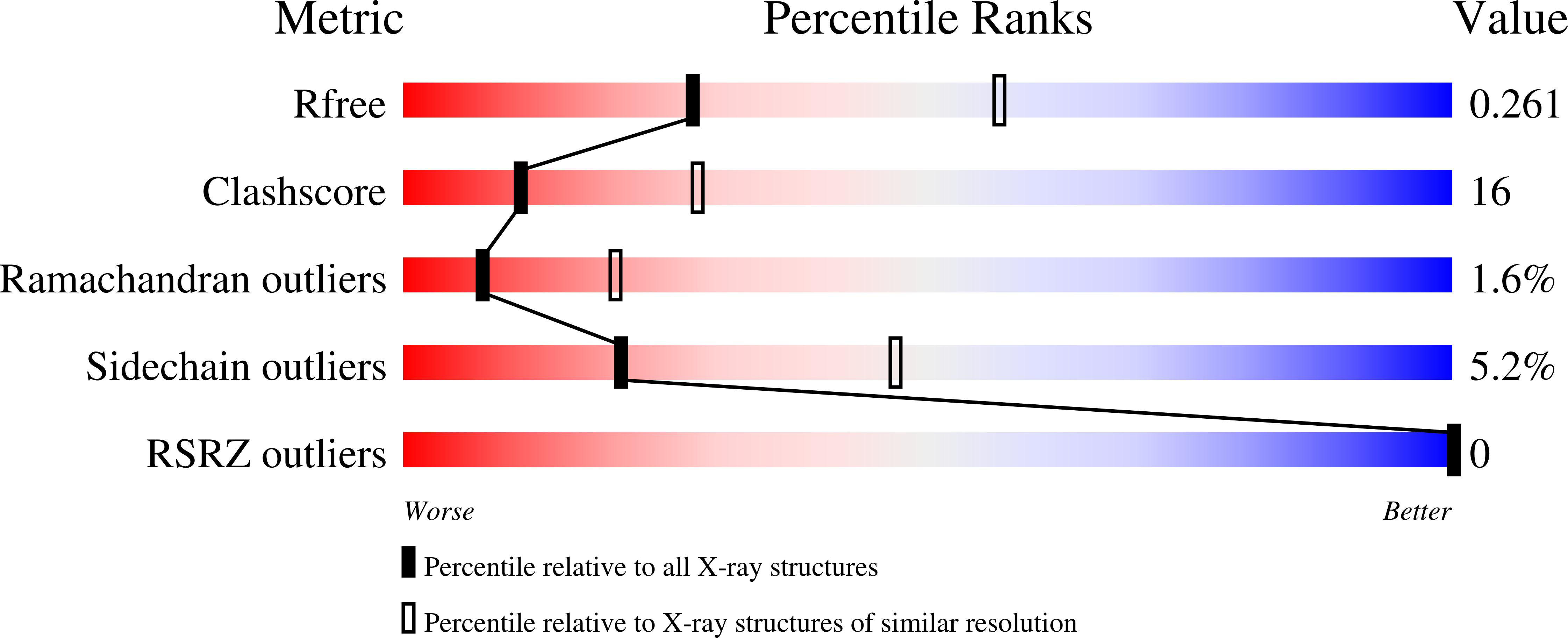
- R-Value Free: 0.251 (Depositor), 0.261 (DCC)
- R-Value Work: 0.191 (Depositor), 0.207 (DCC)
- R-Value Observed: 0.191 (Depositor)
| Length ( Å ) | Angle ( ˚ ) |
|---|---|
| a = 189.26 | α = 90 |
| b = 38.95 | β = 105.46 |
| c = 95.39 | γ = 90 |
| Software Name | Purpose |
|---|---|
| CNS | refinement |
| MOSFLM | data reduction |
| SCALA | data scaling |
| CNS | phasing |
Entry History
Deposition Data
- Released Date: 2010-11-10 Deposition Author(s): Ghuman, J., Curry, S.
Revision History (Full details and data files)
- Version 1.0: 2010-11-10
Type: Initial release - Version 1.1: 2011-05-08
Changes: Version format compliance - Version 1.2: 2011-07-13
Changes: Version format compliance - Version 1.3: 2023-12-20
Changes: Data collection, Database references, Derived calculations, Other, Refinement description - Version 1.4: 2024-10-23
Changes: Structure summary
Best viewed with Netscape Navigator 4.7 or Internet Explorer 6 at 800×600. No cookies required.