GeezerVison is showing www.rcsb.org — RCSB PDB - 3B9L: Human serum albumin complexed with myristate and AZT
3B9L | pdb_00003b9lHuman serum albumin complexed with myristate and AZT
This is version 1.4 of the entry. See complete history. A new drug binding subsite on human serum albumin and drug-drug interaction studied by X-ray crystallographyZhu, L., Yang, F., Chen, L., Meehan, E.J., Huang, M.(2008) J Struct Biol 162: 40-49 - PubMed: 18258455 Search on PubMed
- DOI: https://doi.org/10.1016/j.jsb.2007.12.004
- Primary Citation Related Structures:
3B9L, 3B9M
- PubMed Abstract:
3'-Azido-3'-deoxythymidine (AZT) is the first clinically effective drug for the treatment of human immunodeficiency virus infection. The drug interaction with human serum albumin (HSA) has been an important component in understanding its mechanism of action, especially in drug distribution and in drug-drug interaction on HSA in the case of multi-drug therapy. We present here crystal structures of a ternary HSA-Myr-AZT complex and a quaternary HSA-Myr-AZT-SAL complex (Myr, myristate; SAL, salicylic acid). From this study, a new drug binding subsite on HSA Sudlow site 1 was identified. The presence of fatty acid is needed for the creation of this subsite due to fatty acid induced conformational changes of HSA. Thus, the Sudlow site 1 of HSA can be divided into three non-overlapped subsites: a SAL subsite, an indomethacin subsite and an AZT subsite. Binding of a drug to HSA often influences simultaneous binding of other drugs. From the HSA-Myr-AZT-SAL complex structure, we observed the coexistence of two drugs (AZT and SAL) in Sudlow site 1 and the competition between these two drugs in subdomain IB. These results provide new structural information on HSA-drug interaction and drug-drug interaction on HSA.
Organizational Affiliation: - State Key Laboratory of Structural Chemistry, Fujian Institute of Research on the Structure of Matter, The Chinese Academy of Sciences, 155 Yang Qiao Xi Lu, Fuzhou, Fujian 350002, China. [email protected]
Macromolecule Content - Total Structure Weight: 68.7 kDa
- Atom Count: 4,780
- Modeled Residue Count: 582
- Deposited Residue Count: 585
- Unique protein chains: 1
Small Molecules| Ligands 2 Unique |
|---|
| ID | Chains | Name / Formula / InChI Key | 2D Diagram | 3D Interactions |
|---|
AZZ
Query on AZZ
Download:Ideal Coordinates CCD File
| I [auth A],
J [auth A] | 3'-azido-3'-deoxythymidine
C10 H13 N5 O4
HBOMLICNUCNMMY-XLPZGREQSA-N |  | | MYR
Query on MYR
Download:Ideal Coordinates CCD File
- SDF format, chain B [auth A]
- SDF format, chain C [auth A]
- SDF format, chain D [auth A]
- SDF format, chain E [auth A]
- SDF format, chain G [auth A]
- SDF format, chain H [auth A]
- SDF format, chain F [auth A]
- MOL2 format, chain B [auth A]
- MOL2 format, chain C [auth A]
- MOL2 format, chain D [auth A]
- MOL2 format, chain E [auth A]
- MOL2 format, chain G [auth A]
- MOL2 format, chain H [auth A]
- MOL2 format, chain F [auth A]
- mmCIF format, chain B [auth A]
- mmCIF format, chain C [auth A]
- mmCIF format, chain D [auth A]
- mmCIF format, chain E [auth A]
- mmCIF format, chain G [auth A]
- mmCIF format, chain H [auth A]
- mmCIF format, chain F [auth A]
| B [auth A]
C [auth A]
D [auth A]
E [auth A]
F [auth A]
B [auth A],
C [auth A],
D [auth A],
E [auth A],
F [auth A],
G [auth A],
H [auth A] | MYRISTIC ACID
C14 H28 O2
TUNFSRHWOTWDNC-UHFFFAOYSA-N |  | |
Binding Affinity Annotations | | ID | Source | Binding Affinity |
|---|
| AZZ | BindingDB: 3B9L | Kd: 4.40e+4 (nM) from 1 assay(s) |
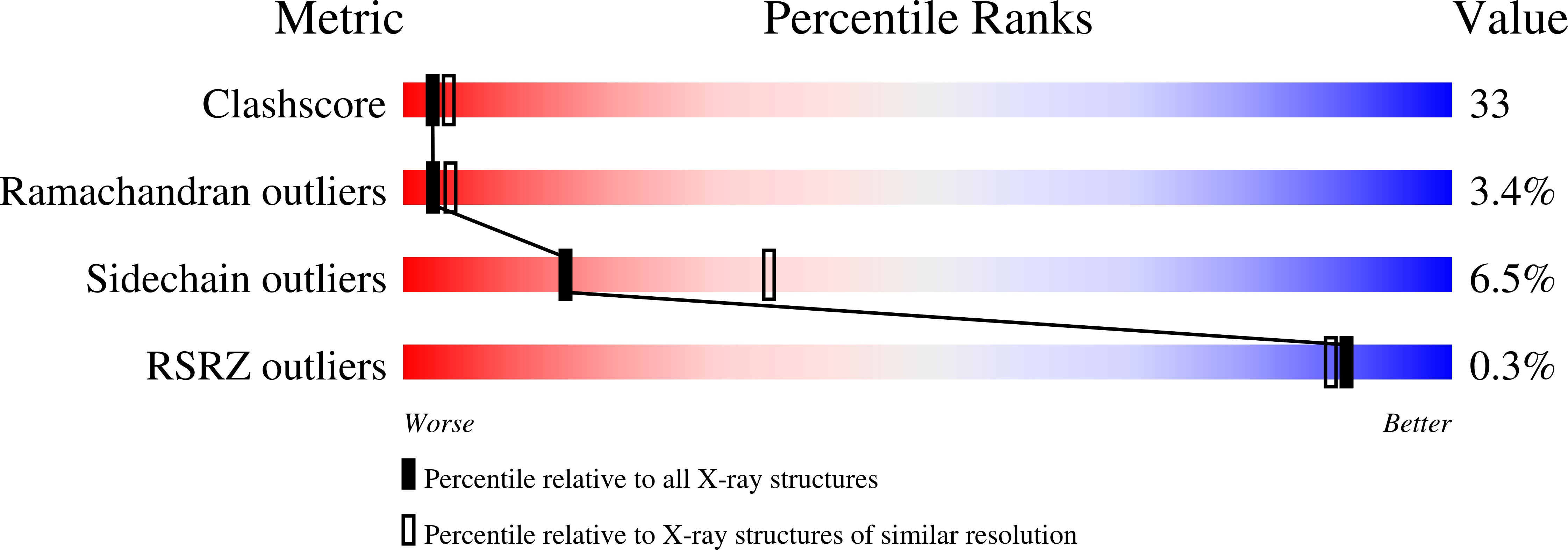
Experimental Data & ValidationExperimental Data- Method: X-RAY DIFFRACTION
- Resolution: 2.60 Å
- R-Value Free: 0.292 (Depositor)
- R-Value Work: 0.223 (Depositor), 0.221 (DCC)
Unit Cell: | Length ( Å ) | Angle ( ˚ ) |
|---|
| a = 183.9 | α = 90 | | b = 38.696 | β = 104.35 | | c = 95.867 | γ = 90 |
| Software Name | Purpose |
|---|
| CNS | refinement | | PDB_EXTRACT | data extraction | | HKL-2000 | data collection | | HKL-2000 | data reduction | | HKL-2000 | data scaling | | CNS | phasing |
Entry History - Version 1.0: 2008-05-27
Type: Initial release
- Version 1.1: 2011-07-13
Changes: Version format compliance - Version 1.2: 2012-07-18
Changes: Non-polymer description - Version 1.3: 2023-11-01
Changes: Data collection, Database references, Derived calculations, Refinement description - Version 1.4: 2024-11-06
Changes: Structure summary
|