GV

GeezerVison

The World Wide Web, restored to 800 pixels of glory.

Home|About|Gaming cabinet|Archives|How it works|Starter sites

On this site

  • Search
  • Advanced Search
  • Sequence Similarity Search
  • 3D Similarity Search
  • Chemical Similarity
  • Browse Annotations
  • New Entries
  • Unreleased Entries
  • PDB Statistics
  • Tools at-a-glance
  • Mol* (MolStar)
  • Sequence Annotations Viewer
  • Sequence Annotations in 3D
  • Genome View

This view

  • Original page
  • New address

GeezerVison is showing www.rcsb.org — RCSB PDB - 3CS7: FACTOR XA IN COMPLEX WITH THE INHIBITOR 1-(4-methoxyphenyl)-6-(4-(1-(pyrrolidin-1-ylmethyl)cyclopropyl)phenyl)-3-(trifluoromethyl)-5,6-dihydro-1H-pyrazolo[3,4-c]pyridin-7(4H)-one

  • Structure Summary
  • Structure
  • Annotations
  • Experiment
  • Sequence
  • Genome
  • Ligands
  • Versions

  • FASTA Sequence
  • mmCIF Format
  • mmCIF Format (Header)
  • Legacy PDB Format
  • Legacy PDB Format (Header)
  • FASTA Sequence
  • PDBx/mmCIF Format
  • PDBx/mmCIF Format (gz)
  • BinaryCIF Format (gz)
  • Legacy PDB Format
  • Legacy PDB Format (gz)
  • PDBML/XML Format (gz)
  • Structure Factors (CIF)
  • Structure Factors (CIF - gz)
  • Validation Full (PDF - gz)
  • Validation (XML - gz)
  • Validation (CIF - gz)
  • Validation 2fo-fc coefficients (CIF - gz)
  • Validation fo-fc coefficients (CIF - gz)
  • Biological Assembly 1 (CIF - gz) 
  • Biological Assembly 1 (PDB - gz)
 Data API 

 3CS7 | pdb_00003cs7

FACTOR XA IN COMPLEX WITH THE INHIBITOR 1-(4-methoxyphenyl)-6-(4-(1-(pyrrolidin-1-ylmethyl)cyclopropyl)phenyl)-3-(trifluoromethyl)-5,6-dihydro-1H-pyrazolo[3,4-c]pyridin-7(4H)-one

  • PDB DOI: https://doi.org/10.2210/pdb3CS7/pdb
  • Classification: HYDROLASE
  • Organism(s): Homo sapiens
  • Mutation(s): No 

  • Deposited: 2008-04-09 Released: 2008-07-08 
  • Deposition Author(s): Alexander, R.S.

Experimental Data Snapshot

  • Method: X-RAY DIFFRACTION
  • Resolution: 2.20 Å
  • R-Value Free: 
    0.274 (Depositor), 0.258 (DCC) 
  • R-Value Work: 
    0.222 (Depositor), 0.209 (DCC) 
  • R-Value Observed: 
    0.225 (Depositor) 

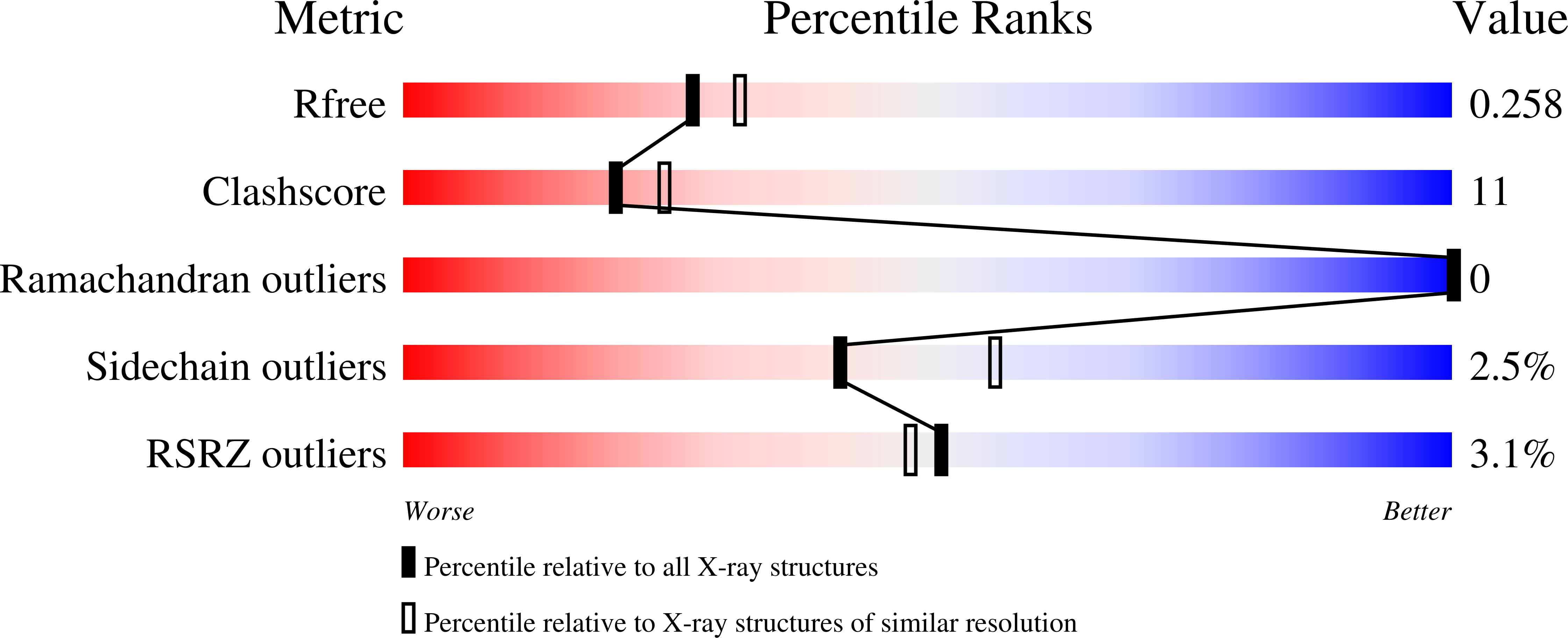
wwPDB Validation 3D Report Full Report

Validation slider image for 3CS7

Ligand Structure Quality Assessment 


This is version 1.2 of the entry. See complete history. 

Literature

  • Download Mendeley

Achieving structural diversity using the perpendicular conformation of alpha-substituted phenylcyclopropanes to mimic the bioactive conformation of ortho-substituted biphenyl P4 moieties: discovery of novel, highly potent inhibitors of Factor Xa.

Qiao, J.X., Cheney, D.L., Alexander, R.S., Smallwood, A.M., King, S.R., He, K., Rendina, A.R., Luettgen, J.M., Knabb, R.M., Wexler, R.R., Lam, P.Y.

(2008) Bioorg Med Chem Lett 18: 4118-4123

  • PubMed: 18550370 Search on PubMed
  • DOI: https://doi.org/10.1016/j.bmcl.2008.05.095
  • Primary Citation Related Structures: 
    3CS7

  • PubMed Abstract: 

    Ortho-substituted biphenyl moieties are widely used in drug design. We herein report a successful use of the perpendicular conformation of the alpha-substituted phenylcyclopropyl groups to mimic the aplanar, biologically active conformation of the ortho-substituted biphenyl moieties to achieve structural diversity. This is exemplified by the design and synthesis of a series of highly potent pyrazole bicyclic-based Factor Xa (FXa) inhibitors bearing alpha-substituted phenylcyclopropyl P4 moieties. The designed perpendicular conformation was confirmed by the X-ray structure of FXa-bound compound 2r. The potential structural basis for the high FXa potency in the phenylcyclopropyl P4 analogs and their improved FXa inhibitory activities compared with the biphenyl P4 counterparts are discussed.


  • Organizational Affiliation: 
    • Bristol-Myers Squibb Company, Research and Development, PO Box 5400, Princeton, NJ 08543-5400, USA. [email protected]
Asymmetric Unit

 Explore in 3D: Structure | Sequence Annotations | Electron Density | Validation Report | Ligand Interaction (LG0)

Biological Assembly 1  

 Explore in 3D: Structure | Sequence Annotations | Electron Density | Validation Report | Ligand Interaction (LG0)


Global Symmetry: Asymmetric - C1 
Global Stoichiometry: Hetero 2-mer - A1B1 


Find Similar Assemblies

Biological assembly 1 assigned by authors and generated by PISA (software)

Macromolecule Content 

  • Total Structure Weight: 32.59 kDa 
  • Atom Count: 2,395 
  • Modeled Residue Count: 286 
  • Deposited Residue Count: 286 
  • Unique protein chains: 2

Macromolecules

Find similar proteins by:
  • 100%
  • 95%
  • 90%
  • 80%
  • 70%
  • 60%
  • 50%
  • 40%
  • 30%
|  3D Structure
Entity ID: 1
MoleculeChains  Sequence LengthOrganismDetailsImage
Coagulation factor X
A
234Homo sapiensMutation(s): 0 
EC: 3.4.21.6
UniProt & NIH Common Fund Data Resources
Find proteins for P00742 (Homo sapiens)
Explore P00742 
Go to UniProtKB:  P00742
GTEx:  ENSG00000126218 
Entity Groups
Sequence Clusters30% Identity50% Identity70% Identity90% Identity95% Identity100% Identity
UniProt GroupP00742
Sequence Annotations
Expand
Reference Sequence
Find similar proteins by:
  • 100%
  • 95%
  • 90%
  • 80%
  • 70%
  • 60%
  • 50%
  • 40%
  • 30%
|  3D Structure
Entity ID: 2
MoleculeChains  Sequence LengthOrganismDetailsImage
Coagulation factor XB [auth L]52Homo sapiensMutation(s): 0 
EC: 3.4.21.6
UniProt & NIH Common Fund Data Resources
Find proteins for P00742 (Homo sapiens)
Explore P00742 
Go to UniProtKB:  P00742
GTEx:  ENSG00000126218 
Entity Groups
Sequence Clusters30% Identity50% Identity70% Identity90% Identity95% Identity100% Identity
UniProt GroupP00742
Sequence Annotations
Expand
Reference Sequence

Small Molecules

Ligands 2 Unique
IDChains Name / Formula / InChI Key2D Diagram3D Interactions
LG0

Query on LG0


Download:Ideal Coordinates CCD File
  • SDF format, chain D [auth A]
  • MOL2 format, chain D [auth A]
  • mmCIF format, chain D [auth A]
D [auth A]1-(4-methoxyphenyl)-6-(4-(1-(pyrrolidin-1-ylmethyl)cyclopropyl)phenyl)-3-(trifluoromethyl)-5,6-dihydro-1H-pyrazolo[3,4-c]pyridin-7(4H)-one
C28 H29 F3 N4 O2
VNHRSGCBTKUBAX-UHFFFAOYSA-N
Interactions
  • Focus chain D [auth A]
Interactions & Density
  • Focus chain D [auth A]
CA

Query on CA


Download:Ideal Coordinates CCD File
  • SDF format, chain C [auth A]
  • MOL2 format, chain C [auth A]
  • mmCIF format, chain C [auth A]
C [auth A]CALCIUM ION
Ca
BHPQYMZQTOCNFJ-UHFFFAOYSA-N
Interactions
  • Focus chain C [auth A]
Interactions & Density
  • Focus chain C [auth A]
Binding Affinity Annotations 
IDSourceBinding Affinity
LG0 BindingDB:  3CS7 Ki: min: 0.02, max: 0.02 (nM) from 2 assay(s)

Experimental Data & Validation

Experimental Data

  • Method: X-RAY DIFFRACTION
  • Resolution: 2.20 Å
  • R-Value Free:  0.274 (Depositor), 0.258 (DCC) 
  • R-Value Work:  0.222 (Depositor), 0.209 (DCC) 
  • R-Value Observed: 0.225 (Depositor) 
Space Group: P 21 21 21
Unit Cell:
Length ( Å )Angle ( ˚ )
a = 56.5α = 90
b = 72.6β = 90
c = 79γ = 90
Software Package:
Software NamePurpose
CNSrefinement
PDB_EXTRACTdata extraction
HKL-2000data reduction
HKL-2000data scaling
CNXrefinement

Structure Validation

View Full Validation Report


Ligand Structure Quality Assessment 


View more in-depth experimental data

Entry History 

Deposition Data

  • Released Date: 2008-07-08 
  • Deposition Author(s): Alexander, R.S.

Revision History  (Full details and data files)

  • Version 1.0: 2008-07-08
    Type: Initial release
  • Version 1.1: 2011-07-13
    Changes: Version format compliance
  • Version 1.2: 2024-10-09
    Changes: Data collection, Database references, Derived calculations, Structure summary
You are visitor 01402503 — GeezerVison does not store the pages you look up.
Best viewed with Netscape Navigator 4.7 or Internet Explorer 6 at 800×600. No cookies required.