GV

GeezerVison

The World Wide Web, restored to 800 pixels of glory.

Home|About|Gaming cabinet|Archives|How it works|Starter sites

On this site

  • Search
  • Advanced Search
  • Sequence Similarity Search
  • 3D Similarity Search
  • Chemical Similarity
  • Browse Annotations
  • New Entries
  • Unreleased Entries
  • PDB Statistics
  • Tools at-a-glance
  • Mol* (MolStar)
  • Sequence Annotations Viewer
  • Sequence Annotations in 3D
  • Genome View

This view

  • Original page
  • New address

GeezerVison is showing www.rcsb.org — RCSB PDB - 4AF3: Human Aurora B Kinase in complex with INCENP and VX-680

  • Structure Summary
  • Structure
  • Annotations
  • Experiment
  • Sequence
  • Genome
  • Ligands
  • Versions

  • FASTA Sequence
  • mmCIF Format
  • mmCIF Format (Header)
  • Legacy PDB Format
  • Legacy PDB Format (Header)
  • FASTA Sequence
  • PDBx/mmCIF Format
  • PDBx/mmCIF Format (gz)
  • BinaryCIF Format (gz)
  • Legacy PDB Format
  • Legacy PDB Format (gz)
  • PDBML/XML Format (gz)
  • Structure Factors (CIF)
  • Structure Factors (CIF - gz)
  • Validation Full (PDF - gz)
  • Validation (XML - gz)
  • Validation (CIF - gz)
  • Validation 2fo-fc coefficients (CIF - gz)
  • Validation fo-fc coefficients (CIF - gz)
  • Biological Assembly 1 (CIF - gz) 
  • Biological Assembly 1 (PDB - gz)
 Data API 

 4AF3 | pdb_00004af3

Human Aurora B Kinase in complex with INCENP and VX-680

  • PDB DOI: https://doi.org/10.2210/pdb4AF3/pdb
  • Classification: TRANSFERASE/INHIBITOR
  • Organism(s): Homo sapiens
  • Expression System: Escherichia coli BL21(DE3)
  • Mutation(s): Yes 

  • Deposited: 2012-01-16 Released: 2012-04-11 
  • Deposition Author(s): Elkins, J.M., Vollmar, M., Wang, J., Picaud, S., Arrowsmith, C.H., Edwards, A., Bountra, C., von Delft, F., Knapp, S.

Experimental Data Snapshot

  • Method: X-RAY DIFFRACTION
  • Resolution: 2.75 Å
  • R-Value Free: 
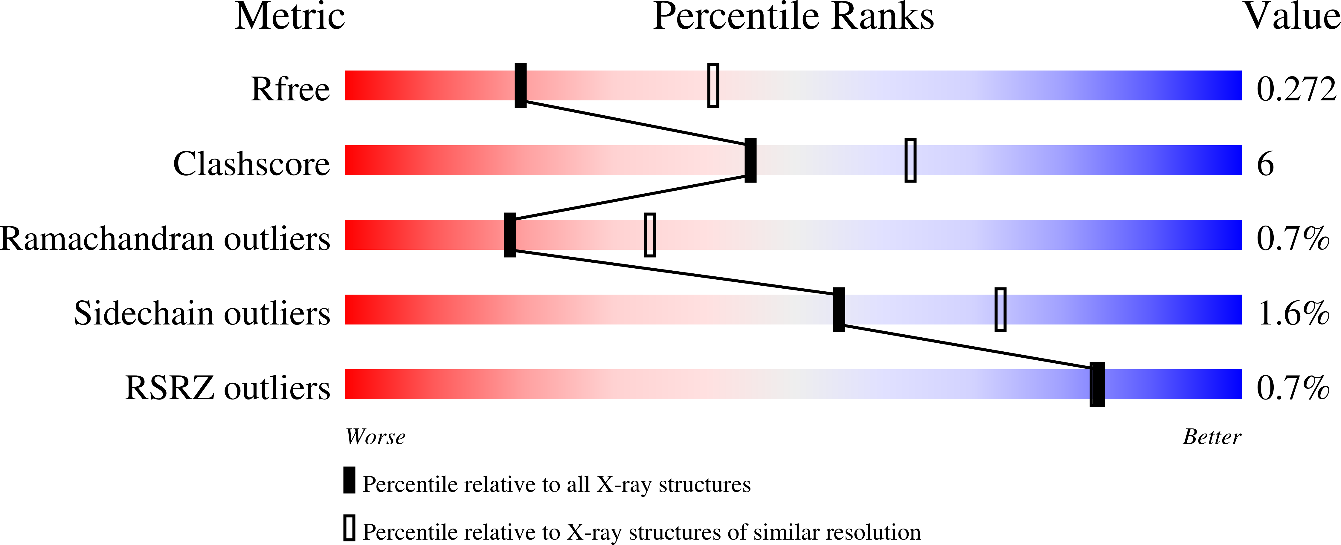
    0.264 (Depositor), 0.272 (DCC) 
  • R-Value Work: 
    0.205 (Depositor), 0.207 (DCC) 
  • R-Value Observed: 
    0.208 (Depositor) 

Starting Model: experimental
View more details

wwPDB Validation 3D Report Full Report

Validation slider image for 4AF3

Ligand Structure Quality Assessment 


This is version 1.3 of the entry. See complete history. 

Literature

  • Download Mendeley

Crystal Structure of Human Aurora B in Complex with Incenp and Vx-680.

Elkins, J.M., Santaguida, S., Musacchio, A., Knapp, S.

(2012) J Med Chem 55: 7841

  • PubMed: 22920039 Search on PubMedSearch on PubMed Central
  • DOI: https://doi.org/10.1021/jm3008954
  • Primary Citation Related Structures: 
    4AF3

  • PubMed Abstract: 

    We present the structure of the human Aurora B kinase domain in complex with the C-terminal Aurora-binding region of human INCENP and the Aurora kinase inhibitor VX-680. The structure unexpectedly reveals a dimeric arrangement of the Aurora B:INCENP complex, which was confirmed to exist in solution by analytical ultracentrifugation. The dimerization involves a domain swap of the activation loop, resulting in a different conformation of the DFG motif as compared to that seen in other kinase complexes with VX-680. The binding of INCENP differs significantly from that seen in the Xenopus laevis Aurora B:INCENP complex currently used as a model for structure-based design for this important oncology target.


  • Organizational Affiliation: 
    • Nuffield Department of Clinical Medicine, Structural Genomics Consortium, University of Oxford , Old Road Campus Research Building, Roosevelt Drive, Oxford, OX3 7DQ, United Kingdom. [email protected]
Asymmetric Unit

 Explore in 3D: Structure | Sequence Annotations | Electron Density | Validation Report | Ligand Interaction (VX6)

Biological Assembly 1  

 Explore in 3D: Structure | Sequence Annotations | Electron Density | Validation Report | Ligand Interaction (VX6)


Global Symmetry: Asymmetric - C1 
Global Stoichiometry: Hetero 2-mer - A1B1 


Find Similar Assemblies

Biological assembly 1 assigned by authors and generated by PISA (software)

Macromolecule Content 

  • Total Structure Weight: 42.4 kDa 
  • Atom Count: 2,427 
  • Modeled Residue Count: 296 
  • Deposited Residue Count: 363 
  • Unique protein chains: 2

Macromolecules

Find similar proteins by:
  • 100%
  • 95%
  • 90%
  • 80%
  • 70%
  • 60%
  • 50%
  • 40%
  • 30%
|  3D Structure
Entity ID: 1
MoleculeChains  Sequence LengthOrganismDetailsImage
AURORA KINASE B
A
292Homo sapiensMutation(s): 1 
EC: 2.7.11.1
UniProt & NIH Common Fund Data Resources
Find proteins for Q96GD4 (Homo sapiens)
Explore Q96GD4 
Go to UniProtKB:  Q96GD4
GTEx:  ENSG00000178999 
Entity Groups
Sequence Clusters30% Identity50% Identity70% Identity90% Identity95% Identity100% Identity
UniProt GroupQ96GD4
Sequence Annotations
Expand
Reference Sequence
Find similar proteins by:
  • 100%
  • 95%
  • 90%
  • 80%
  • 70%
  • 60%
  • 50%
  • 40%
  • 30%
|  3D Structure
Entity ID: 2
MoleculeChains  Sequence LengthOrganismDetailsImage
INNER CENTROMERE PROTEINB [auth D]71Homo sapiensMutation(s): 0 
UniProt & NIH Common Fund Data Resources
Find proteins for Q9NQS7 (Homo sapiens)
Explore Q9NQS7 
Go to UniProtKB:  Q9NQS7
PHAROS:  Q9NQS7
GTEx:  ENSG00000149503 
Entity Groups
Sequence Clusters30% Identity50% Identity70% Identity90% Identity95% Identity100% Identity
UniProt GroupQ9NQS7
Sequence Annotations
Expand
Reference Sequence

Small Molecules

Ligands 1 Unique
IDChains Name / Formula / InChI Key2D Diagram3D Interactions
VX6

Query on VX6


Download:Ideal Coordinates CCD File
  • SDF format, chain C [auth A]
  • MOL2 format, chain C [auth A]
  • mmCIF format, chain C [auth A]
C [auth A]CYCLOPROPANECARBOXYLIC ACID {4-[4-(4-METHYL-PIPERAZIN-1-YL)-6-(5-METHYL-2H-PYRAZOL-3-YLAMINO)-PYRIMIDIN-2-YLSULFANYL]-PHENYL}-AMIDE
C23 H28 N8 O S
GCIKSSRWRFVXBI-UHFFFAOYSA-N
Interactions
  • Focus chain C [auth A]
Interactions & Density
  • Focus chain C [auth A]

Experimental Data & Validation

Experimental Data

  • Method: X-RAY DIFFRACTION
  • Resolution: 2.75 Å
  • R-Value Free:  0.264 (Depositor), 0.272 (DCC) 
  • R-Value Work:  0.205 (Depositor), 0.207 (DCC) 
  • R-Value Observed: 0.208 (Depositor) 
Space Group: P 31 2 1
Unit Cell:
Length ( Å )Angle ( ˚ )
a = 80.145α = 90
b = 80.145β = 90
c = 92.542γ = 120
Software Package:
Software NamePurpose
REFMACrefinement
XDSdata reduction
Aimlessdata scaling
SAWAYAdata scaling
PHASERphasing

Structure Validation

View Full Validation Report


Ligand Structure Quality Assessment 


View more in-depth experimental data

Entry History 

Deposition Data

  • Released Date: 2012-04-11 
  • Deposition Author(s): Elkins, J.M., Vollmar, M., Wang, J., Picaud, S., Arrowsmith, C.H., Edwards, A., Bountra, C., von Delft, F., Knapp, S.

Revision History  (Full details and data files)

  • Version 1.0: 2012-04-11
    Type: Initial release
  • Version 1.1: 2012-09-26
    Changes: Database references, Structure summary
  • Version 1.2: 2018-01-24
    Changes: Structure summary
  • Version 1.3: 2023-12-20
    Changes: Data collection, Database references, Derived calculations, Other, Refinement description
You are visitor 01402503 — GeezerVison does not store the pages you look up.
Best viewed with Netscape Navigator 4.7 or Internet Explorer 6 at 800×600. No cookies required.