GV

GeezerVison

The World Wide Web, restored to 800 pixels of glory.

Home|About|Gaming cabinet|Archives|How it works|Starter sites

On this site

  • Search
  • Advanced Search
  • Sequence Similarity Search
  • 3D Similarity Search
  • Chemical Similarity
  • Browse Annotations
  • New Entries
  • Unreleased Entries
  • PDB Statistics
  • Tools at-a-glance
  • Mol* (MolStar)
  • Sequence Annotations Viewer
  • Sequence Annotations in 3D
  • Genome View

This view

  • Original page
  • New address

GeezerVison is showing www.rcsb.org — RCSB PDB - 4FVG: SPFH domain of mouse stomatin (Crystal form 3)

  • Structure Summary
  • Structure
  • Annotations
  • Experiment
  • Sequence
  • Genome
  • Versions

  • FASTA Sequence
  • mmCIF Format
  • mmCIF Format (Header)
  • Legacy PDB Format
  • Legacy PDB Format (Header)
  • FASTA Sequence
  • PDBx/mmCIF Format
  • PDBx/mmCIF Format (gz)
  • BinaryCIF Format (gz)
  • Legacy PDB Format
  • Legacy PDB Format (gz)
  • PDBML/XML Format (gz)
  • Structure Factors (CIF)
  • Structure Factors (CIF - gz)
  • Validation Full (PDF - gz)
  • Validation (XML - gz)
  • Validation (CIF - gz)
  • Validation 2fo-fc coefficients (CIF - gz)
  • Validation fo-fc coefficients (CIF - gz)
  • Biological Assembly 1 (CIF - gz) 
  • Biological Assembly 1 (PDB - gz)
 Data API 

 4FVG | pdb_00004fvg

SPFH domain of mouse stomatin (Crystal form 3)

  • PDB DOI: https://doi.org/10.2210/pdb4FVG/pdb
  • Classification: MEMBRANE PROTEIN
  • Organism(s): Mus musculus
  • Expression System: Escherichia coli
  • Mutation(s): Yes 

  • Deposited: 2012-06-29 Released: 2012-08-15 
  • Deposition Author(s): Brand, J., Schwefel, D., Daumke, O.

Experimental Data Snapshot

  • Method: X-RAY DIFFRACTION
  • Resolution: 1.80 Å
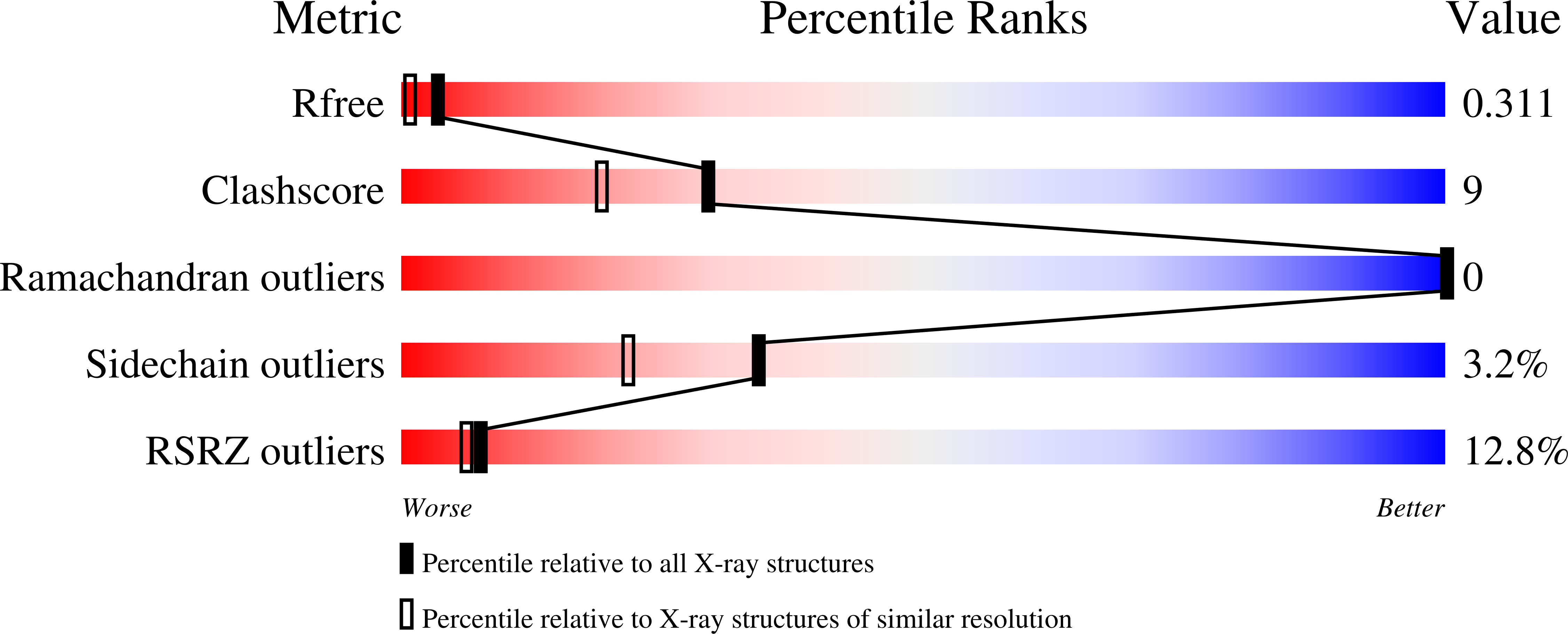
  • R-Value Free: 
    0.245 (Depositor), 0.311 (DCC) 
  • R-Value Work: 
    0.194 (Depositor), 0.268 (DCC) 
  • R-Value Observed: 
    0.196 (Depositor) 

Starting Model: experimental
View more details

wwPDB Validation 3D Report Full Report

Validation slider image for 4FVG

This is version 1.2 of the entry. See complete history. 

Literature

  • Download Mendeley

A stomatin dimer modulates the activity of acid-sensing ion channels.

Brand, J., Smith, E.S., Schwefel, D., Lapatsina, L., Poole, K., Omerbasic, D., Kozlenkov, A., Behlke, J., Lewin, G.R., Daumke, O.

(2012) EMBO J 31: 3635-3646

  • PubMed: 22850675 Search on PubMedSearch on PubMed Central
  • DOI: https://doi.org/10.1038/emboj.2012.203
  • Primary Citation Related Structures: 
    4FVF, 4FVG, 4FVJ

  • PubMed Abstract: 

    Stomatin proteins oligomerize at membranes and have been implicated in ion channel regulation and membrane trafficking. To obtain mechanistic insights into their function, we determined three crystal structures of the conserved stomatin domain of mouse stomatin that assembles into a banana-shaped dimer. We show that dimerization is crucial for the repression of acid-sensing ion channel 3 (ASIC3) activity. A hydrophobic pocket at the inside of the concave surface is open in the presence of an internal peptide ligand and closes in the absence of this ligand, and we demonstrate a function of this pocket in the inhibition of ASIC3 activity. In one crystal form, stomatin assembles via two conserved surfaces into a cylindrical oligomer, and these oligomerization surfaces are also essential for the inhibition of ASIC3-mediated currents. The assembly mode of stomatin uncovered in this study might serve as a model to understand oligomerization processes of related membrane-remodelling proteins, such as flotillin and prohibitin.


  • Organizational Affiliation: 
    • Max-Delbrück Center for Molecular Medicine, Crystallography Department, Berlin, Germany.
Asymmetric Unit

 Explore in 3D: Structure | Sequence Annotations | Electron Density | Validation Report | Ligand Interaction (CD)

Biological Assembly 1  

 Explore in 3D: Structure | Sequence Annotations | Electron Density | Validation Report | Ligand Interaction (CD)


Global Symmetry: Cyclic - C2  (Explore in 3D)
Global Stoichiometry: Homo 2-mer - A2 


Find Similar Assemblies

Biological assembly 1 assigned by authors and generated by PISA (software)

Macromolecule Content 

  • Total Structure Weight: 14.64 kDa 
  • Atom Count: 969 
  • Modeled Residue Count: 109 
  • Deposited Residue Count: 133 
  • Unique protein chains: 1

Macromolecules

Find similar proteins by:
  • 100%
  • 95%
  • 90%
  • 80%
  • 70%
  • 60%
  • 50%
  • 40%
  • 30%
|  3D Structure
Entity ID: 1
MoleculeChains  Sequence LengthOrganismDetailsImage
Stomatin
A
133Mus musculusMutation(s): 3 
Gene Names: Epb7.2, Epb72, Stom
UniProt
Find proteins for P54116 (Mus musculus)
Explore P54116 
Go to UniProtKB:  P54116
Entity Groups
Sequence Clusters30% Identity50% Identity70% Identity90% Identity95% Identity100% Identity
UniProt GroupP54116
Sequence Annotations
Expand
Reference Sequence

Small Molecules

Ligands 1 Unique
IDChains Name / Formula / InChI Key2D Diagram3D Interactions
CD

Query on CD


Download:Ideal Coordinates CCD File
  • SDF format, chain C [auth A]
  • SDF format, chain B [auth A]
  • MOL2 format, chain C [auth A]
  • MOL2 format, chain B [auth A]
  • mmCIF format, chain C [auth A]
  • mmCIF format, chain B [auth A]
B [auth A],
C [auth A]
CADMIUM ION
Cd
WLZRMCYVCSSEQC-UHFFFAOYSA-N
Interactions
  • Focus chain B [auth A]
  • Focus chain C [auth A]
Interactions & Density
  • Focus chain B [auth A]
  • Focus chain C [auth A]

Experimental Data & Validation

Experimental Data

  • Method: X-RAY DIFFRACTION
  • Resolution: 1.80 Å
  • R-Value Free:  0.245 (Depositor), 0.311 (DCC) 
  • R-Value Work:  0.194 (Depositor), 0.268 (DCC) 
  • R-Value Observed: 0.196 (Depositor) 
Space Group: P 64 2 2
Unit Cell:
Length ( Å )Angle ( ˚ )
a = 84.489α = 90
b = 84.489β = 90
c = 68.937γ = 120
Software Package:
Software NamePurpose
XDSdata scaling
MOLREPphasing
REFMACrefinement
XDSdata reduction

Structure Validation

View Full Validation Report


View more in-depth experimental data

Entry History 

Deposition Data

  • Released Date: 2012-08-15 
  • Deposition Author(s): Brand, J., Schwefel, D., Daumke, O.

Revision History  (Full details and data files)

  • Version 1.0: 2012-08-15
    Type: Initial release
  • Version 1.1: 2012-10-31
    Changes: Database references
  • Version 1.2: 2023-09-13
    Changes: Data collection, Database references, Derived calculations, Refinement description
You are visitor 01402503 — GeezerVison does not store the pages you look up.
Best viewed with Netscape Navigator 4.7 or Internet Explorer 6 at 800×600. No cookies required.