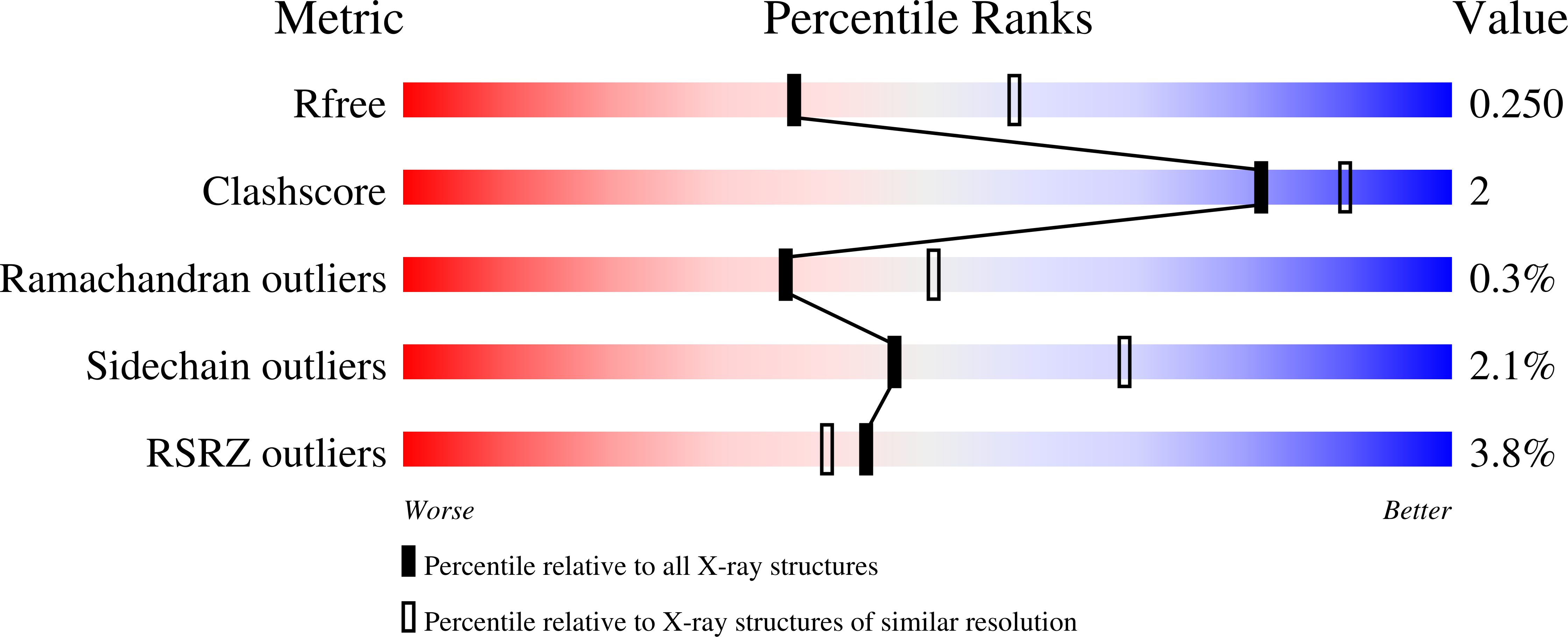
GeezerVison is showing www.rcsb.org — RCSB PDB - 4K71: Crystal structure of a high affinity Human Serum Albumin variant bound to the Neonatal Fc Receptor
4K71 | pdb_00004k71Crystal structure of a high affinity Human Serum Albumin variant bound to the Neonatal Fc Receptor - Classification: LIPID TRANSPORT
- Organism(s): Homo sapiens
- Expression System: Saccharomyces cerevisiae, Homo sapiens
- Mutation(s): Yes
- Deposited: 2013-04-16 Released: 2013-10-23
- Deposition Author(s): Schmidt, M.M., Townson, S.A., Andreucci, A., Dombrowski, C., Erbe, D.V., King, B., Kovalchin, J.T., Masci, A., Murillo, A., Schirmer, E.B., Furfine, E.S., Barnes, T.M.
Experimental Data Snapshot - Method: X-RAY DIFFRACTION
- Resolution: 2.40 Å
- R-Value Free:
0.255 (Depositor), 0.250 (DCC) - R-Value Work:
0.212 (Depositor), 0.212 (DCC) - R-Value Observed:
0.214 (Depositor)
This is version 1.2 of the entry. See complete history. Crystal structure of an HSA/FcRn complex reveals recycling by competitive mimicry of HSA ligands at a pH-dependent hydrophobic interface.Schmidt, M.M., Townson, S.A., Andreucci, A.J., King, B.M., Schirmer, E.B., Murillo, A.J., Dombrowski, C., Tisdale, A.W., Lowden, P.A., Masci, A.L., Kovalchin, J.T., Erbe, D.V., Wittrup, K.D., Furfine, E.S., Barnes, T.M.(2013) Structure 21: 1966-1978 - PubMed: 24120761 Search on PubMed
- DOI: https://doi.org/10.1016/j.str.2013.08.022
- Primary Citation Related Structures:
4K71
- PubMed Abstract:
The long circulating half-life of serum albumin, the most abundant protein in mammalian plasma, derives from pH-dependent endosomal salvage from degradation, mediated by the neonatal Fc receptor (FcRn). Using yeast display, we identified human serum albumin (HSA) variants with increased affinity for human FcRn at endosomal pH, enabling us to solve the crystal structure of a variant HSA/FcRn complex. We find an extensive, primarily hydrophobic interface stabilized by hydrogen-bonding networks involving protonated histidines internal to each protein. The interface features two key FcRn tryptophan side chains inserting into deep hydrophobic pockets on HSA that overlap albumin ligand binding sites. We find that fatty acids (FAs) compete with FcRn, revealing a clash between ligand binding and recycling, and that our high-affinity HSA variants have significantly increased circulating half-lives in mice and monkeys. These observations open the way for the creation of biotherapeutics with significantly improved pharmacokinetics.
Organizational Affiliation: - Eleven Biotherapeutics, Inc., Cambridge, MA 02142, USA.
Macromolecule Content - Total Structure Weight: 219.08 kDa
- Atom Count: 15,259
- Modeled Residue Count: 1,888
- Deposited Residue Count: 1,916
- Unique protein chains: 3
Small Molecules| Ligands 1 Unique |
|---|
| ID | Chains | Name / Formula / InChI Key | 2D Diagram | 3D Interactions |
|---|
SO4
Query on SO4
Download:Ideal Coordinates CCD File
- SDF format, chain G [auth A]
- SDF format, chain H [auth A]
- SDF format, chain I [auth A]
- SDF format, chain J [auth A]
- SDF format, chain K [auth A]
- SDF format, chain L [auth A]
- SDF format, chain M [auth A]
- SDF format, chain N [auth B]
- SDF format, chain O [auth B]
- SDF format, chain P [auth C]
- SDF format, chain Q [auth C]
- SDF format, chain S [auth D]
- SDF format, chain T [auth D]
- SDF format, chain U [auth D]
- SDF format, chain V [auth D]
- SDF format, chain W [auth D]
- SDF format, chain X [auth E]
- SDF format, chain Y [auth F]
- SDF format, chain R [auth D]
- MOL2 format, chain G [auth A]
- MOL2 format, chain H [auth A]
- MOL2 format, chain I [auth A]
- MOL2 format, chain J [auth A]
- MOL2 format, chain K [auth A]
- MOL2 format, chain L [auth A]
- MOL2 format, chain M [auth A]
- MOL2 format, chain N [auth B]
- MOL2 format, chain O [auth B]
- MOL2 format, chain P [auth C]
- MOL2 format, chain Q [auth C]
- MOL2 format, chain S [auth D]
- MOL2 format, chain T [auth D]
- MOL2 format, chain U [auth D]
- MOL2 format, chain V [auth D]
- MOL2 format, chain W [auth D]
- MOL2 format, chain X [auth E]
- MOL2 format, chain Y [auth F]
- MOL2 format, chain R [auth D]
- mmCIF format, chain G [auth A]
- mmCIF format, chain H [auth A]
- mmCIF format, chain I [auth A]
- mmCIF format, chain J [auth A]
- mmCIF format, chain K [auth A]
- mmCIF format, chain L [auth A]
- mmCIF format, chain M [auth A]
- mmCIF format, chain N [auth B]
- mmCIF format, chain O [auth B]
- mmCIF format, chain P [auth C]
- mmCIF format, chain Q [auth C]
- mmCIF format, chain S [auth D]
- mmCIF format, chain T [auth D]
- mmCIF format, chain U [auth D]
- mmCIF format, chain V [auth D]
- mmCIF format, chain W [auth D]
- mmCIF format, chain X [auth E]
- mmCIF format, chain Y [auth F]
- mmCIF format, chain R [auth D]
| G [auth A]
H [auth A]
I [auth A]
J [auth A]
K [auth A]
G [auth A],
H [auth A],
I [auth A],
J [auth A],
K [auth A],
L [auth A],
M [auth A],
N [auth B],
O [auth B],
P [auth C],
Q [auth C],
R [auth D],
S [auth D],
T [auth D],
U [auth D],
V [auth D],
W [auth D],
X [auth E],
Y [auth F] | SULFATE ION
O4 S
QAOWNCQODCNURD-UHFFFAOYSA-L |  | |
Experimental Data & ValidationExperimental Data- Method: X-RAY DIFFRACTION
- Resolution: 2.40 Å
- R-Value Free: 0.255 (Depositor), 0.250 (DCC)
- R-Value Work: 0.212 (Depositor), 0.212 (DCC)
- R-Value Observed: 0.214 (Depositor)
Unit Cell: | Length ( Å ) | Angle ( ˚ ) |
|---|
| a = 127.893 | α = 90 | | b = 203.536 | β = 90 | | c = 100.561 | γ = 90 |
| Software Name | Purpose |
|---|
| HKL-2000 | data collection | | PHASER | phasing | | REFMAC | refinement | | XDS | data reduction | | XSCALE | data scaling |
Entry History Deposition Data- Released Date: 2013-10-23
Deposition Author(s): Schmidt, M.M., Townson, S.A., Andreucci, A., Dombrowski, C., Erbe, D.V., King, B., Kovalchin, J.T., Masci, A., Murillo, A., Schirmer, E.B., Furfine, E.S., Barnes, T.M.
- Version 1.0: 2013-10-23
Type: Initial release
- Version 1.1: 2014-02-05
Changes: Database references - Version 1.2: 2024-11-27
Changes: Data collection, Database references, Derived calculations, Structure summary
|