GeezerVisonThe World Wide Web, restored to 800 pixels of glory. | |||||||||||||||||||||||||||||||||||||||||||||||||||||||||||||||||||||||||||||||||||||||||||||||||||||||||||
| Home|About|Gaming cabinet|Archives|How it works|Starter sites | |||||||||||||||||||||||||||||||||||||||||||||||||||||||||||||||||||||||||||||||||||||||||||||||||||||||||||
GeezerVison is showing www.rcsb.org — RCSB PDB - 4PZI: Zinc finger region of MLL2 in complex with CpG DNA
| |||||||||||||||||||||||||||||||||||||||||||||||||||||||||||||||||||||||||||||||||||||||||||||||||||||||||||
Entity ID: 1 | |||||
|---|---|---|---|---|---|
| Molecule | Chains | Sequence Length | Organism | Details | Image |
| Histone-lysine N-methyltransferase 2B | 67 | Homo sapiens | Mutation(s): 0 Gene Names: KMT2B, HRX2, KIAA0304, MLL2, MLL4, TRX2, WBP7 EC: 2.1.1.43 (PDB Primary Data), 2.1.1.364 (UniProt) |  | |
UniProt & NIH Common Fund Data Resources | |||||
PHAROS: Q9UMN6 GTEx: ENSG00000272333 | |||||
Entity Groups | |||||
| Sequence Clusters | 30% Identity50% Identity70% Identity90% Identity95% Identity100% Identity | ||||
| UniProt Group | Q9UMN6 | ||||
Sequence AnnotationsExpand | |||||
Reference Sequence | |||||

Small Molecules
| Ligands 2 Unique | |||||
|---|---|---|---|---|---|
| ID | Chains | Name / Formula / InChI Key | 2D Diagram | 3D Interactions | |
| ZN Download:Ideal Coordinates CCD File | D [auth A], E [auth A] | ZINC ION Zn PTFCDOFLOPIGGS-UHFFFAOYSA-N |  | Interactions Interactions & Density | |
| UNX Download:Ideal Coordinates CCD File | F [auth A] | UNKNOWN ATOM OR ION X |  | Interactions Interactions & Density | |
Experimental Data & Validation
Experimental Data
- Method: X-RAY DIFFRACTION
- Resolution: 2.15 Å
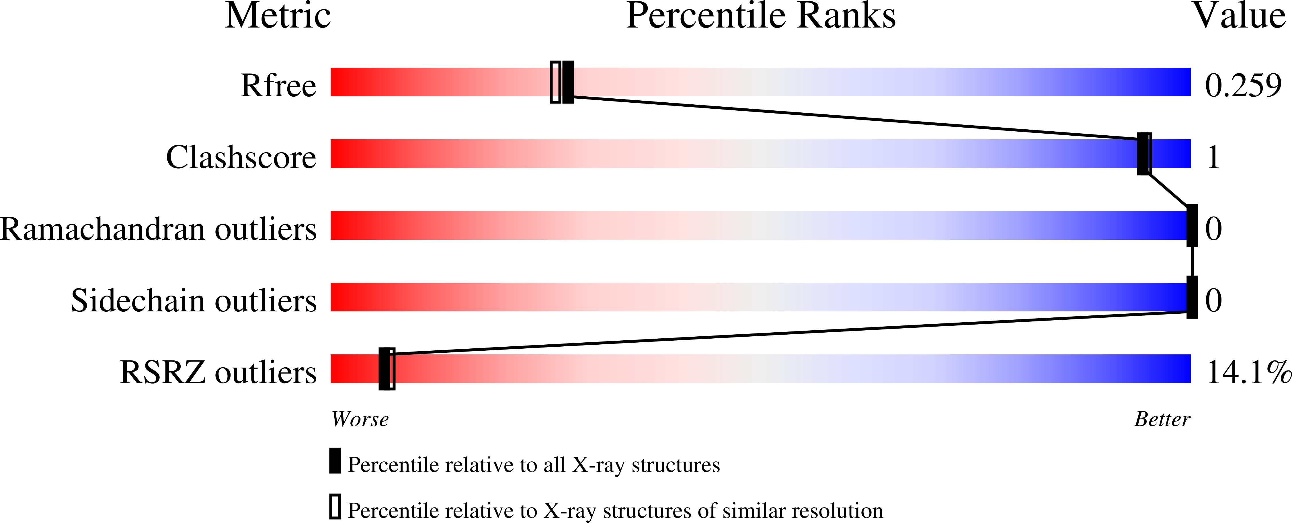
- R-Value Free: 0.273 (Depositor), 0.259 (DCC)
- R-Value Work: 0.226 (Depositor)
- R-Value Observed: 0.228 (Depositor)
| Length ( Å ) | Angle ( ˚ ) |
|---|---|
| a = 70.736 | α = 90 |
| b = 41.005 | β = 99.24 |
| c = 55.508 | γ = 90 |
| Software Name | Purpose |
|---|---|
| Aimless | data scaling |
| PHASER | phasing |
| REFMAC | refinement |
| PDB_EXTRACT | data extraction |
| XDS | data reduction |
Entry History
Deposition Data
- Released Date: 2014-06-04 Deposition Author(s): Chao, X., Tempel, W., Liu, K., Dong, A., Bountra, C., Weigelt, J., Arrowsmith, C.H., Edwards, A.M., Min, J., Structural Genomics Consortium (SGC)
Revision History (Full details and data files)
- Version 1.0: 2014-06-04
Type: Initial release - Version 1.1: 2018-01-03
Changes: Database references - Version 1.2: 2022-08-24
Changes: Database references, Derived calculations - Version 1.3: 2024-02-28
Changes: Data collection - Version 1.4: 2024-04-03
Changes: Refinement description
Best viewed with Netscape Navigator 4.7 or Internet Explorer 6 at 800×600. No cookies required.