GV

GeezerVison

The World Wide Web, restored to 800 pixels of glory.

Home|About|Gaming cabinet|Archives|How it works|Starter sites

On this site

  • Search
  • Advanced Search
  • Sequence Similarity Search
  • 3D Similarity Search
  • Chemical Similarity
  • Browse Annotations
  • New Entries
  • Unreleased Entries
  • PDB Statistics
  • Tools at-a-glance
  • Mol* (MolStar)
  • Sequence Annotations Viewer
  • Sequence Annotations in 3D
  • Genome View

This view

  • Original page
  • New address

GeezerVison is showing www.rcsb.org — RCSB PDB - 2GZK: Structure of a complex of tandem HMG boxes and DNA

  • Structure Summary
  • Structure
  • Annotations
  • Experiment
  • Sequence
  • Genome
  • Versions

  • FASTA Sequence
  • mmCIF Format
  • mmCIF Format (Header)
  • Legacy PDB Format
  • Legacy PDB Format (Header)
  • FASTA Sequence
  • PDBx/mmCIF Format
  • PDBx/mmCIF Format (gz)
  • BinaryCIF Format (gz)
  • Legacy PDB Format
  • Legacy PDB Format (gz)
  • PDBML/XML Format (gz)
  • Validation Full (PDF - gz)
  • Validation (XML - gz)
  • Validation (CIF - gz)
  • Biological Assembly 1 (CIF - gz) 
  • Biological Assembly 1 (PDB - gz)
 Data API 

 2GZK | pdb_00002gzk

Structure of a complex of tandem HMG boxes and DNA

  • PDB DOI: https://doi.org/10.2210/pdb2GZK/pdb
  • NAKB: 2GZK
  • Classification: DNA/STRUCTURAL PROTEIN
  • Organism(s): Homo sapiens, Rattus rattus
  • Expression System: Escherichia coli
  • Mutation(s): No 

  • Deposited: 2006-05-11 Released: 2006-07-25 
  • Deposition Author(s): Stott, K., Tang, G.S., Lee, K.B., Thomas, J.O.

Experimental Data Snapshot

  • Method: SOLUTION NMR
  • Conformers Calculated: 100 
  • Conformers Submitted: 1 
  • Selection Criteria: structures with the lowest energy 

wwPDB Validation 3D Report Full Report

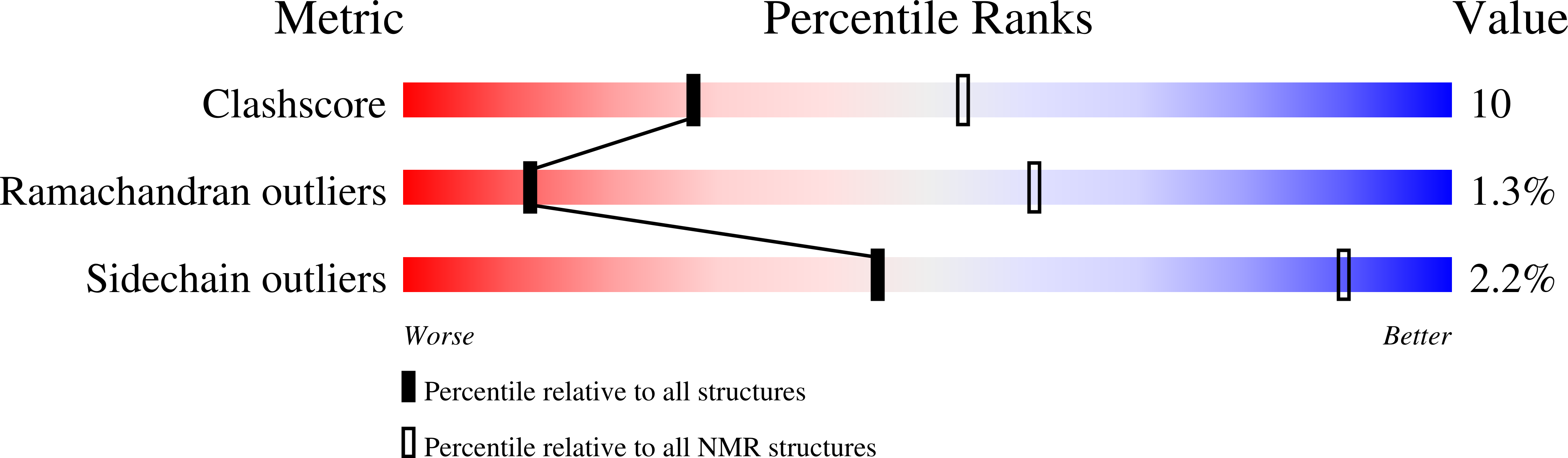
Validation slider image for 2GZK

This is version 1.4 of the entry. See complete history. 

Literature

  • Download Mendeley

Structure of a Complex of Tandem HMG Boxes and DNA.

Stott, K., Tang, G.S., Lee, K.B., Thomas, J.O.

(2006) J Mol Biol 360: 90-104

  • PubMed: 16813837 Search on PubMed
  • DOI: https://doi.org/10.1016/j.jmb.2006.04.059
  • Primary Citation Related Structures: 
    2GZK

  • PubMed Abstract: 

    The high-mobility group protein HMGB1 contains two tandem DNA-binding HMG box domains, A and B, linked by a short flexible linker that allows the two domains to behave independently in the free protein. There is no structural information on how the linked domains and linker behave when bound to DNA, mainly due to the lack of any DNA-sequence preference of HMGB1. We report the structure determination, by NMR spectroscopy, of a well-defined complex of two tandem HMG boxes bound to a 16 bp oligonucleotide. The protein is an engineered version of the AB di-domain of HMGB1, in which the A box has been replaced by the HMG box of the sequence-specific transcription factor SRY, to give SRY.B. In the SRY.B/DNA complex, both HMG boxes bind in the minor groove and contribute to the overall DNA bending by intercalation of bulky hydrophobic residues between base-pairs; the bends reinforce each other, and the basic linker lies partly in the minor groove. As well as being the first structure of an HMG-box di-domain bound to DNA, this provides the first structure of the B domain of HMGB1 bound to DNA.


  • Organizational Affiliation: 
    • Department of Biochemistry, University of Cambridge, 80 Tennis Court Road, Cambridge CB2 1GA, UK.
NMR Ensemble

 Explore in 3D: Structure | Sequence Annotations | Validation Report

Biological Assembly 1  

 Explore in 3D: Structure | Sequence Annotations | Validation Report


Global Symmetry: Asymmetric - C1 
Global Stoichiometry: Monomer - A1 


Find Similar Assemblies

Biological assembly 1 assigned by authors.

Macromolecule Content 

  • Total Structure Weight: 28.78 kDa 
  • Atom Count: 1,986 
  • Modeled Residue Count: 191 
  • Deposited Residue Count: 191 
  • Unique protein chains: 1
  • Unique nucleic acid chains: 2

Macromolecules


Find similar proteins by:
  • 100%
  • 95%
  • 90%
  • 80%
  • 70%
  • 60%
  • 50%
  • 40%
  • 30%
|  3D Structure
Entity ID: 3
MoleculeChains  Sequence LengthOrganismDetailsImage
Sex-determining region on Y / HMGB1C [auth A]159Homo sapiens, Rattus rattus
This entity is chimeric
Mutation(s): 0 
UniProt & NIH Common Fund Data Resources
Find proteins for P63159 (Rattus norvegicus)
Explore P63159 
Go to UniProtKB:  P63159
Find proteins for Q05066 (Homo sapiens)
Explore Q05066 
Go to UniProtKB:  Q05066
GTEx:  ENSG00000184895 
Entity Groups
Sequence Clusters30% Identity50% Identity70% Identity90% Identity95% Identity100% Identity
UniProt GroupsP63159Q05066
Sequence Annotations
Expand
Reference Sequence
Find similar nucleic acids by:  Sequence
Entity ID: 1
MoleculeChains LengthOrganismImage
5'-D(*GP*GP*GP*AP*TP*CP*TP*AP*AP*AP*CP*AP*AP*TP*GP*C)-3'A [auth B]16N/A
Sequence Annotations
Expand
Reference Sequence
Find similar nucleic acids by:  Sequence
Entity ID: 2
MoleculeChains LengthOrganismImage
5'-D(*GP*CP*AP*TP*TP*GP*TP*TP*TP*AP*GP*AP*TP*CP*CP*C)-3'B [auth C]16N/A
Sequence Annotations
Expand
Reference Sequence

Experimental Data & Validation

Experimental Data

  • Method: SOLUTION NMR
  • Conformers Calculated: 100 
  • Conformers Submitted: 1 
  • Selection Criteria: structures with the lowest energy 

Structure Validation

View Full Validation Report


View more in-depth experimental data

Entry History 

Deposition Data

  • Released Date: 2006-07-25 
  • Deposition Author(s): Stott, K., Tang, G.S., Lee, K.B., Thomas, J.O.

Revision History  (Full details and data files)

  • Version 1.0: 2006-07-25
    Type: Initial release
  • Version 1.1: 2008-05-01
    Changes: Version format compliance
  • Version 1.2: 2011-07-13
    Changes: Version format compliance
  • Version 1.3: 2022-03-09
    Changes: Data collection, Database references, Derived calculations
  • Version 1.4: 2024-05-29
    Changes: Data collection
You are visitor 01402503 — GeezerVison does not store the pages you look up.
Best viewed with Netscape Navigator 4.7 or Internet Explorer 6 at 800×600. No cookies required.